
Figure 2.
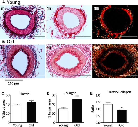
Collagen and elastin content of mesenteric small arteries. (Ai and Bi) Representative sections of mesenteric small arteries (MSA) stained with Miller reagent for elastin, (Aii, Aiii and Bii, Biii) representative sections of MSA stained with picrosirius red for collagen illuminated with (ii) normal and (iii) polarized light. Upper panels MSA from young and lower panels MSA from old animals. Quantitation of (C) elastin, (D) collagen, and (E) elastin/collagen ratio. Data are expressed as mean ± SEM from individual arteries from seven young and seven old animals, *P <0.05 by Student's t‐test.