
Figure 3.
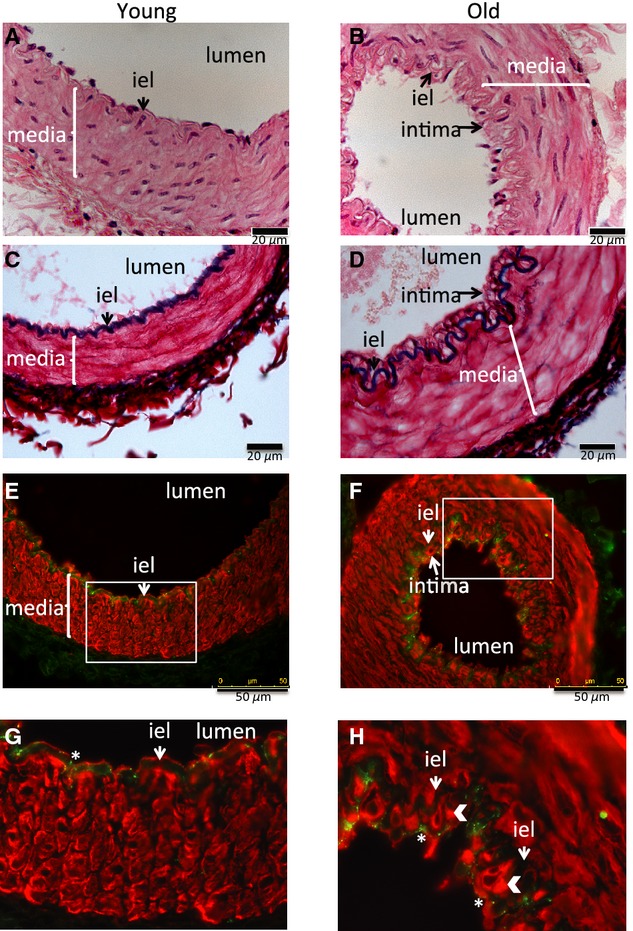
Age‐related intima formation. Representative sections of mesenteric artery stained with hematoxylin and eosin (A and B), Miller reagent for elastin (C and D), von Willebrand Factor (green) for endothelial cells, and α‐smooth muscle actin (red) for smooth muscle cells (E–H) from (A, C, E, and G) young animal and (B, D, F, and H) old animal. (G and H) Enlargement of the areas within the white box in (E and F). White arrow heads in (H) show intimal α‐smooth cell actin‐positive cells, * in G and H show von Willebrand staining in endothelial cells, “iel” internal elastic lamina. A–D scale bar 20 μm, E and F scale bar 50 μm.