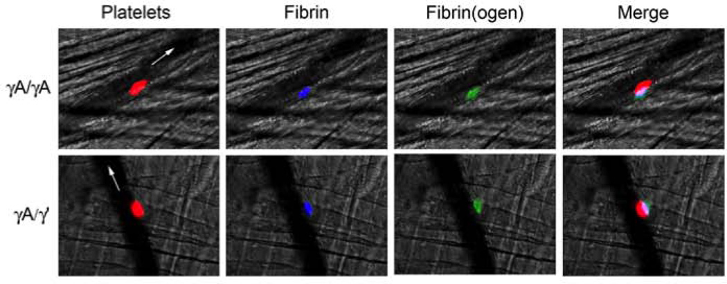
Figure 3. Intravital microscopy shows both γA/γA and γA/γ’ isoforms are incorporated into murine thrombi.
Venules were visualized in the cremaster muscle of mice infused with HBS (control) or AlexaFluor 594-labeled anti-platelet (anti-GPIX) antibody, AlexaFluor 647-labeled anti-fibrin antibody, and purified γA/γA or γA/γ’ directly labeled with AlexaFluor 488. Thrombosis was triggered via laser injury. Flow is indicated by white arrows. Colors are: platelets (red), fibrin(ogen) (green), and fibrin (blue). In the merged image, colors are: platelets plus fibrin(ogen) (pink), platelets plus fibrin (purple), and fibrin(ogen) plus fibrin (teal). Images show representative thrombi from 3–4 mice with 14–20 injuries total.

