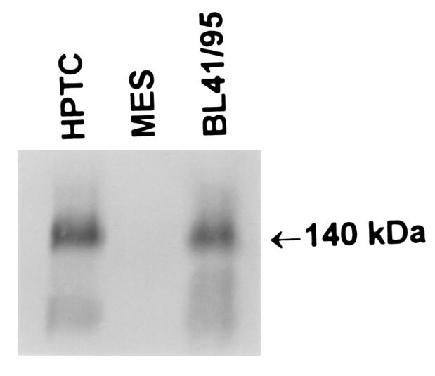
Figure 6.

Detection of CD21 by immunoprecipitation and SDS-PAGE. Cultured human proximal tubule cells (HPTC), human mesangial cells (MES), and EBV-infected Burkitt’s lymphoma cells (BL 41/95) were [35S]methionine labeled. Cells were lysed and immunoprecipitated with the HB5 CD21 mAb, and then lysates were subjected to SDS-PAGE on a 7.5% gel. Dried gels were autoradiographed at –70°C overnight, before development. One of 4 similar autoradiograms is shown.
