
Figure 6.
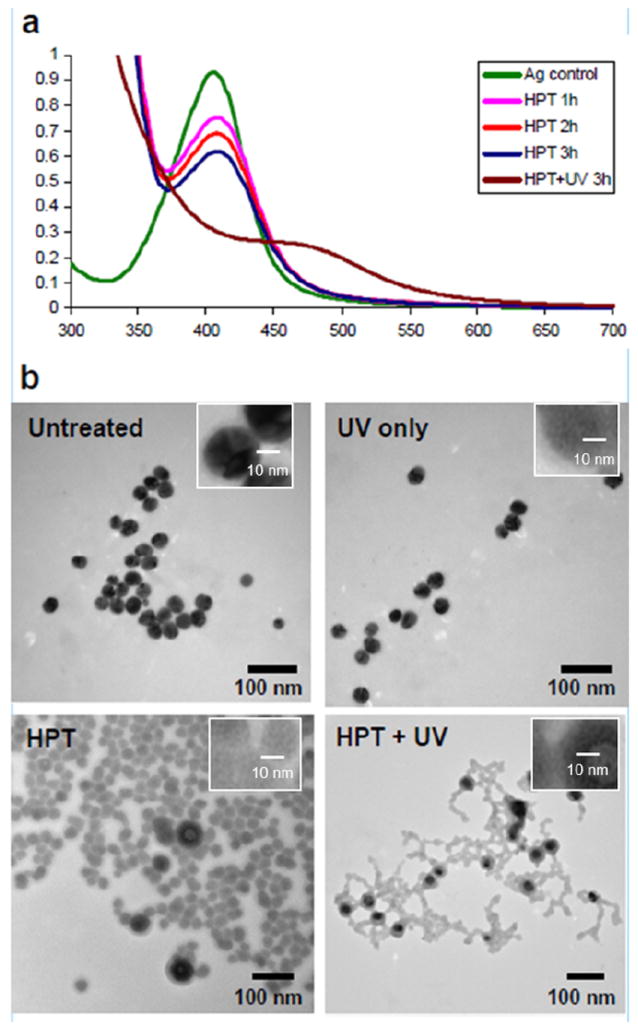
(A) UV-vis spectra of 20 nm silver nanoparticles with treatment of HPT (5 μM), alone or in combination with UV treatment. Addition of UV treatment shows dramatic absorbance differences. (B) TEM images of 20 nm citrate-stabilized silver nanoparticles: untreated (control), treated with UV for 3 hours, treated with HPT (5 μM) for 3 hours, and treated with HPT (5 μM) and UV for 3 hours. From the inserts, it is clear the crystal structure of the silver was destroyed by UV, HPT and HPT + UV treatments.