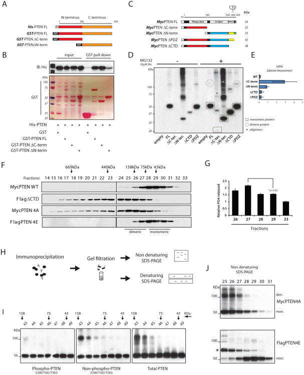
Figure 2. Dimerization defines a pool of catalytically active PTEN.
(A) Diagram of recombinant proteins showing PTEN full length (FL) and deletion mutants. In red, N-terminus domain, amino-acid (a.a.) 1-185; in orange, C-terminus domain, a.a. 186-403.
(B) GST-PTENFL and domains were purified (Ponceau-S staining); His-PTEN pulled down is detected by Western blot.
(C) Schematic representing series of Myc-tagged PTENFL and deletion mutant vectors. Predicted molecular weights are indicated.
(D) PC3 cells were transfected with the indicated expression vectors. Total lysates were resolved by non-reducing SDS-PAGE and probed with an anti-Myc antibody. Circles and squares indicate monomeric and dimeric PTEN conformations, respectively. Asterisks indicate oligomers of PTEN domains.
(E) Ratio between PTEN dimer/monomer in PTEN FL and deletion mutant series. Mean values with associated SD are shown.
(F) Lysates from HEK293 cells transfected with the indicated PTEN vectors were separated by gel filtration. Fractions were resolved by SDS-PAGE and probed with specific tag antibodies.
(G) Fractions containing different conformations of PTEN and collected as in (F) were tested for their activity toward PIP3, and normalized over protein levels. Mean values from triplicate wells with associated SD are shown.
(H) Experimental flow chart: cell lysates were subjected to IP with an anti-Myc antibody. Immuno-complexes were eluted in native conditions and separated by tandem-column gel filtration (See also Figure S2H). Collected fractions were resolved by reducing (Figure S2H) and non-reducing SDS-PAGE (Figure 2I).
(I) Fractions collected as in (H) were resolved by non-reducing SDS-PAGE and probed with the indicated PTEN antibodies.
(J) Non-reducing SDS-PAGE of eluted fractions generated as in (F) were blotted with anti-Myc (top panel) and anti-Flag (bottom panel) antibodies. Asterisk indicates a shift in FlagPTEN4E probably due to post-translational modifications.

