
Figure 1. Locations of the epitopes and their sequences among the NaV subtypes.
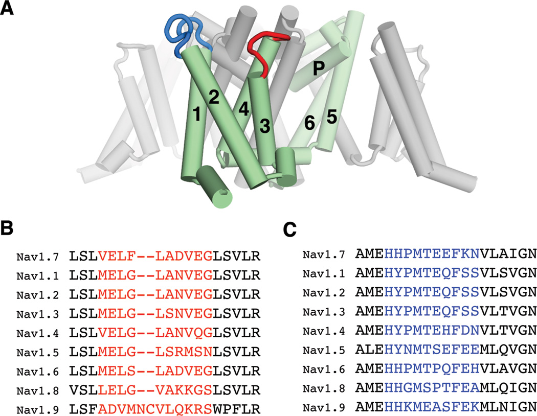
(A) The chosen epitopes are mapped on the crystal structure of a bacterial NaV channel (Payandeh et al., 2011). One of the four repeats, composed of 6 transmembrane helices (S1–S6; 1–6), is colored green. The loops between S3–S4 and between S1–S2 are colored red and blue, respectively. (B) and (C) Sequence alignments corresponding to the S3–S4 (B) and S1–S2 (C) loops of hNaV subtypes. The regions chosen for raising antibodies are colored red and blue, respectively.