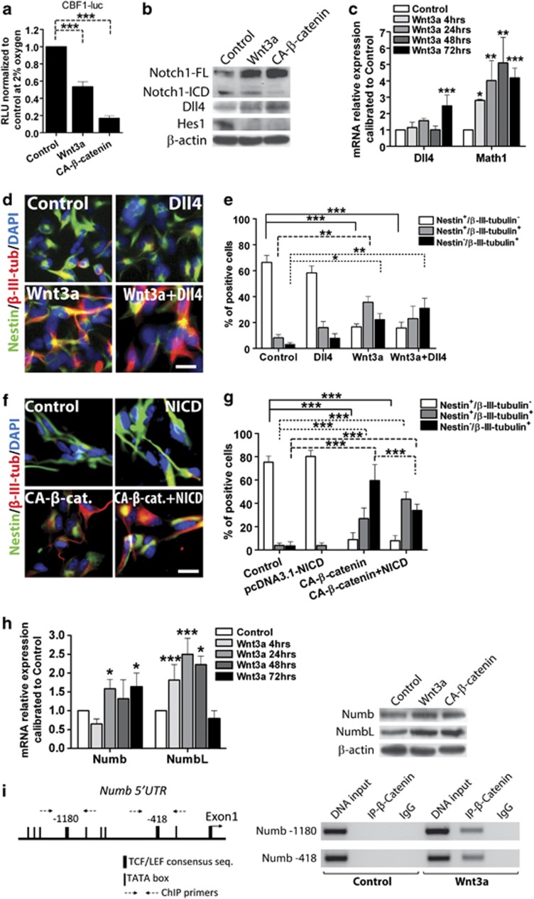
Figure 3.
Wnt pathway activation inhibits Notch signalling in GBM-derived cells. (a) CBF1-luc reporter analysis of Wnt3a-treated or CA-β-catenin-transfected cells at 2% O2. Mean of four tumours±S.E.M., n=2 for each tumour. (b) WB of protein extracts from same cells as in (a) displaying Notch pathway regulation. (c) RQ-PCR analysis reporting relative expression of Dll4 and Math1. Mean of six tumours±S.E.M., n=4 for each tumour. (d and e) Representative immunofluorescence images of GBM cells treated with Dll4, Wnt3a or both for 96 h and stained for Nestin (green)/β-III-tubulin (red) (d) and graph reporting relative quantification (e). Mean of three tumours±S.E.M., n=3 for each tumour. Bar=100 μm. (f and g) Representative immunofluorescence images of GBM cells transfected with NICD, CA-β-catenin or both, cultured for 48 h and stained for Nestin (green)/β-III-tubulin (red) (f) and bar graph reporting relative quantification (g). Mean of three tumours±S.E.M., n=3 for each tumour. Bar=100 μm. (h) RQ-PCR analysis showing mRNA levels of Numb and NumbL of Wnt3a-treated GBM cells at different time points (left). Numb and NumbL protein expression of Wnt3a-treated or CA-β-catenin-transfected GBM cells (right). Mean of six tumours±S.E.M., n=4 for each tumour. (i) ChIP analysis of Numb promoter performed on 293T and GBM cells treated with Wnt3a or not treated. The IP was performed using anti-total β-catenin antibody or an irrelevant antibody as negative control. *P<0.05, **P<0.01, ***P<0.001

