
Extended Data Figure 1. Esr1 mRNA expression in Esr1cre/+ male and female mice.
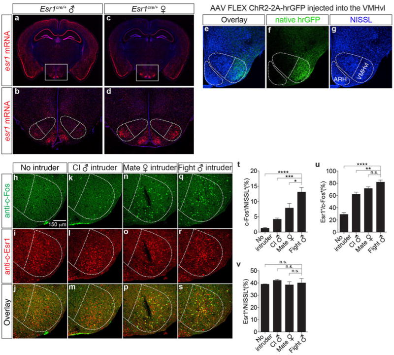
In situ hybridization for Esr1mRNA in Esr1cre/+ male (a, b, red) and female (c, d, red) mice (Bregma ∼-1.65 mm). b-d are the boxed areas in a-c. Note that the expression of esr1 mRNA in VMHvl (dotted outline) is higher in females than in males. e-g. Immunofluorescence showing that expression of a Cre-dependent hrGFP reporter expressed from a stereotaxically injected rAAV (f, green) is restricted to VMHvl, without detectable spillover expression in the nearby arcuate hypothalamic nucleus (ARH). h-s. Double labeling for behaviorally-induced c-Fos (h, k, n, q, anti-c-Fos, green) and Esr1 (i, l, o, r, anti-Esr1, red) in wild-type male residents following a 30-min resident-intruder test with no (h-j, n=3), male (k-m, close investigation without attack, n=4; q-s, attack, n=5) or female (n-p, mating, n=5) intruders. t-v. Quantification of the fraction of total (NISSL+) cells that were c-Fos+ following different behaviors (t), fraction of c-Fos+ that were Esr1+for each behavior (u), and fraction of NISSL+ cells that are Esr1+(v) in VMHvl, quantified from data as illustrated in (h-s). *p<0.05, ***p<0.001, ****p<0.0001; one-way ANOVA with Dunnett's multiple comparisons test.