
Fig. 3. MYSM1 has an intrinsic role in B-cell development.
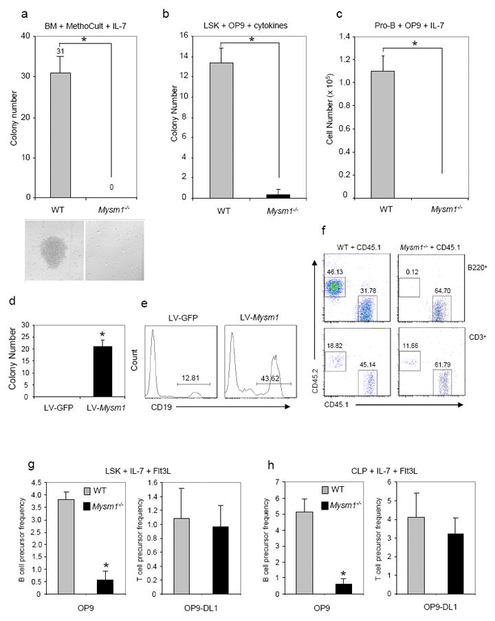
a. Pre-B colony assays of bone marrow cells from Mysm1-/-mice and WT littermates. B-cell colony numbers (n=3) and representative colony morphology (bottom) on day 7 of MethoCult M3630 culture (StemCell Technologies) containing IL-7 are shown from one of three repeated experiments. *P, <0.01, Mysm1-/-vs. WT.
b. B-cell colony formation assays of sorted Lin-c-Kit+Sca1+ (LSK) hematopoietic stem cells on the OP9 stromal cell culture in the presence of SCF, Flt3, and IL-7 for 10 days. B-cell colony numbers per well (n=3) are shown from one of two repeated experiments. *P, <0.01, Mysm1-/-vs. WT.
c. In vitro proliferation of sorted pro-B cells (B220+CD43+IgM-) from the bone marrow on OP9 stromal cells supplemented with IL-7 for 5 days. B-cell numbers per well (n=3) are shown from one of two repeated experiments.
d & e. MYSM1 rescue assays. Bone marrow cells were transduced with a recombinant lentiviral vector LV-Mysm1 or a control LV-GFP. The transduced BM cells were then subject to pre-B colony assays and B-cell colony numbers were scored on day 7 (d). Flow cytometric analysis was performed on day 10 of the culture (e). Data are showed from one of three repeated experiments. *P, < 0.01, LV-Mysm1 vs. LV-GFP.
f. Bone marrow transplantation. A 1:1 mixture of whole BM cells from CD45.2 Mysm1-/-mice or WT littermates and CD45.1 congenic mice were injected retro-orbitally into irradiated (9.5 Gy) CD45.1 congenic mice (2 × 10⁶ cells/mouse, n=6 per group). After 3 wks, the relative percentages of B220+ B-cells or CD3+ T cells expressing donor (CD45.2+) or recipient (CD45.1+) derived population were determined by flow cytometry analysis. Results are representative of two independent experiments.
g & h. B-cell and T-cell colony formation assays of HSC (LSK) (g) and CLP (h) cells sorted from BM of WT and Mysm1-/-mice on the OP9 or OP-DL1 stromal cell culture in the presence of Flt3 and IL-7 for 10 days. B-cell and T-cell colony numbers per 100 seeded WT or Mysm1-/-HSC or CLP cells are shown from one of two repeated experiments. *P, < 0.01, Mysm1-/-vs. WT OP9 culture.