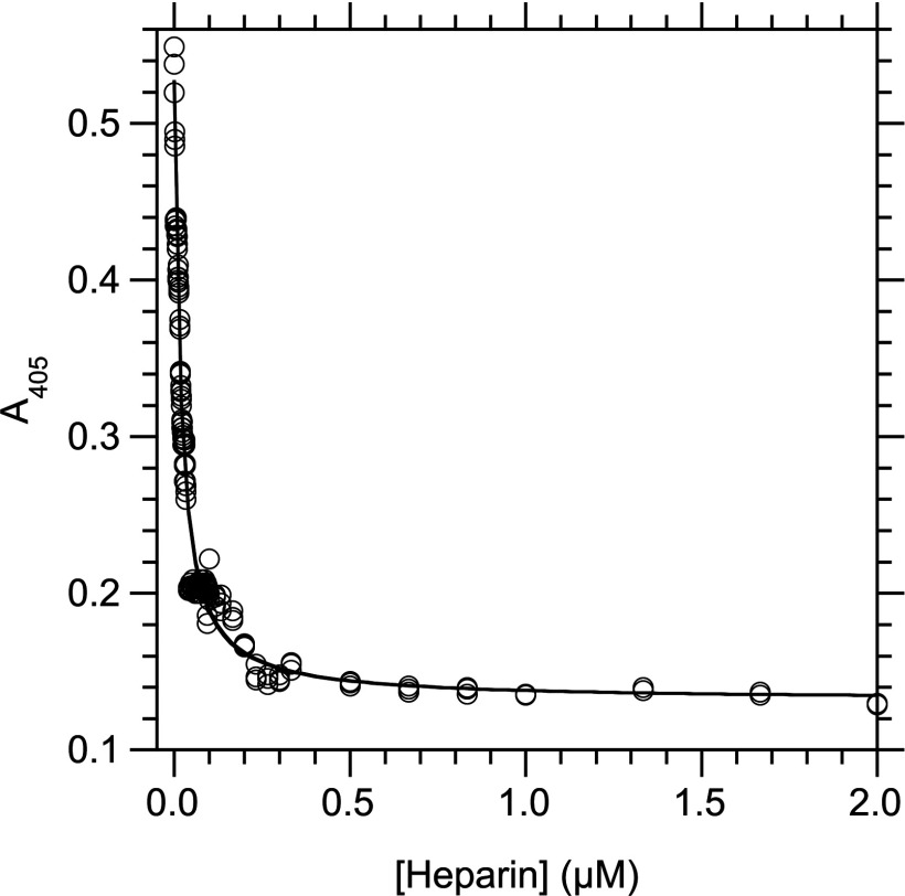
Figure 4. Determination of Ki at 25°C for the inhibition of the interaction between RPSA-(225–295) and laminin by heparin.
Laminin (5 μg/ml, 5.5 nM) and heparin were first incubated for 20 h at 25°C in solution (buffer C) until the binding reaction reached equilibrium. The concentration of free laminin was then measured by an indirect ELISA in which RPSA-(225–295) (0.15 μg/ml, 16 nM) was immobilized in the wells of a microtitre plate and the captured laminin was revealed with a specific antibody. The total concentration of heparin in the binding reaction is given along the x-axis; the A405 signal, which is linearly related to the concentration of free laminin in the binding reaction, is given along the y-axis. The curve was obtained by fitting eqn (5) to the experimental data. Totally, 52 concentrations of heparin were used and each data point was done in triplicate.

