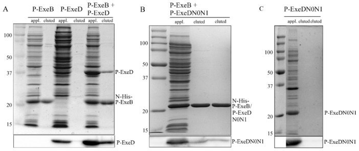
Figure 6. Co-purification of P-ExeD and P-ExeDN0N1 with N-His-P-ExeB.

Cell lysates were applied to a Ni affinity chromatography column and eluted with 0.5(upper panel) or immunoblotted with α-ExeD serum (lower panel). Cell lysates from E. coli expressing either N-His tagged P-ExeB, or P-ExeD are also shown. Cell lysates from E. coli co-expressing untagged P-ExeD and N-His tagged P-ExeB (A), untagged P-ExeDN0N1 and N-His tagged P-ExeB (B) or expressing untagged P-ExeDN0N1 alone (C) were purified and analyzed as described above. The P-ExeDN0N1 and P-ExeB fragments have similar sizes, therefore, in panel B the P-ExeDN0N1 fragment can only be distinguished in the immunoblot.
