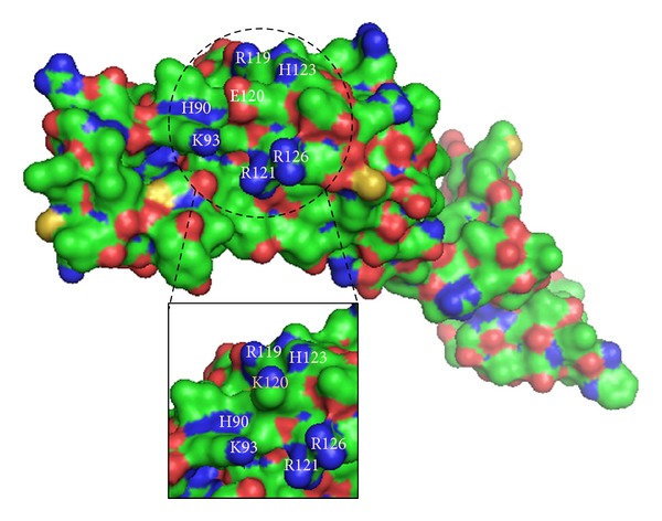
Figure 5.

Bulk representation of wild-type NANOS3, with p.Glu120Lys mutant shown in details box. The dashed circle highlights a protein surface region rich in basic residues, shown in blue. At the center of this region, lies the acidic residue glutamic acid 120 (E120), shown in red. In the details, substitution by lysine 120 (K120) disturbs the electrostatic interactions among adjacent residues.
