
Fig. 1.
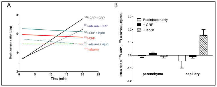
Permeation of 125I-CRP from blood to brain in C57 mice, with 131I-albumin as a reference substance. (A) Multiple-time regression analysis showing the overall influx rate. Trace amounts of CRP did not cross the BBB, shown by a slightly sloping regression line almost parallel to that of albumin. Co-treatment of leptin did not affect the influx rate (slope of the regression line) and the increase of the volume of distribution (y-axis values) was not significant. Co-treatment with CRP initially seemed to depress the volume of distribution of 125I-CRP in the first 10 min, but overall induced an influx of 125I-CRP (Ki = 0.22 ± 0.13 μl/g-min) and 131I-albumin (Ki = 0.16 ± 0.11 μl/g-min). (B) Capillary depletion study showing the distribution of radioactively labeled tracers in cerebral cortical parenchyma and vasculature. The influx rate of 125I-CRP to brain parenchyma, normalized by subtracting the 131I-albumin space, was only positive in the group treated with excess unlabeled CRP. This contrasts with an increased vascular accumulation in the leptin treated group.