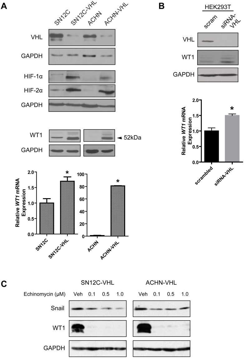
Figure 1. Knockdown of VHL increases WT1 expression.
(A) Confirmation of VHL knockdown (top), and analysis of WT1 protein (middle) and mRNA (bottom) in the isogenic SN12C and ACHN cell lines. (B) HEK293T cells were transfected with scrambled oligoneucleotides or VHL-specific siRNAs and WT1 protein (top) and mRNA (bottom) was measured. Graphs show the mean±SD of one representative of three independent experiments. *, P<0.05. (C) SN12C-VHL (left) and ACHN-VHL (right) cells were treated with vehicle (DMSO) or echinomycin (0.1, 0.5, 1.0 µM) for 24 hr and protein expression was assessed by immunoblot.

