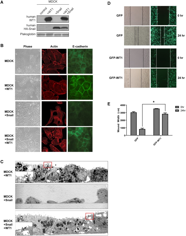
Figure 5. WT1 preserves epithelial junctions and suppresses motility in renal cells.
(A) MDCK cells were transfected as indicated and protein expression was assessed by immunoblot. (B) Representative immunofluorescene images of MDCK cells transfected with Snail alone or Snail and WT1. (C) Electron microscopy images of MDCK cells transfected with WT1, Snail, or WT1 and Snail. Red boxes indicate cell-cell contact regions showing intercellular junctions junctions enlarged in the inset (arrow). Note the absence of intercellular contacts and spindle morphology of Snail-transfected cells. (D) HEK293T cells were transfected with either GFP or GFP-WT1 and a scratch motility assay was performed. (E) Quantification of the width of the wound at the indicated time points. Graph depicts mean±SD of three independent experiments. *, P<0.05.

