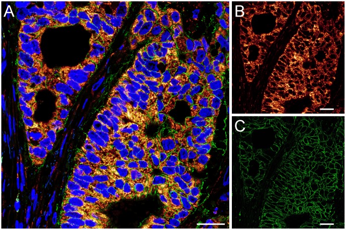
Figure 1. Immunofluorescence labeling of HER2 positive paraffin-embedded rectal cancer tissue.
Confocal overview image of a region of a HER2 positive rectal cancer tissue section labeled with DAPI (blue) to highlight the nuclei and decorated with antisera against Tom20 (fire) and HER2 (green). (A) overlay, (B) Tom20, and (C) HER2. Scale bars: 25 µm.

