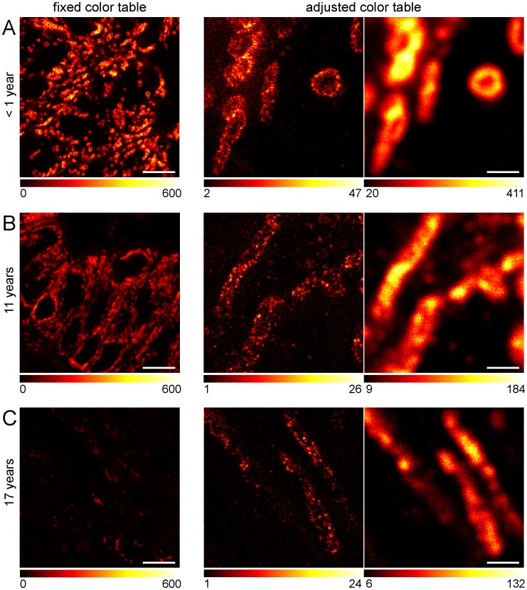
Figure 4. STED super-resolution microscopy of archived human tissue samples stored for up to 17 years in a clinical repository.
Representative images of tumor tissues stored at room temperature for less than 1 year (A), 11 years (B) or 17 years (C), were sectioned, dewaxed, decorated with an antiserum against Tom20 and imaged. Left: Representative confocal images. The same color table was used for the three images in order to visualize the relative staining efficiencies. Middle/Right: Comparison of STED (middle) and confocal (right) microscopy of tissue sections of different age. Here, the color tables were adjusted to the signal intensities obtained. Scale bars: 10 µm (left) and 1 µm (middle, right).

