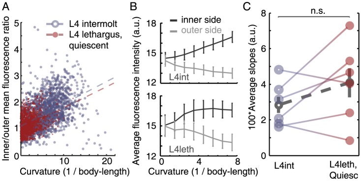
Figure 2. Bends are actively maintained during lethargus quiescence bouts.

(A) Values of curvature at all positions along the body were plotted against the ratio of fluorescence of a calcium reporter expressed in the inner and outer body-wall muscles at the same bend. All points plotted were measured from a single animal during L4int (blue, empty circles) and during L4leth bouts of quiescence (red, filled circles). Dashed lines denote the linear correlations between the curvature and the ratio of fluorescence during L4int (blue) and L4leth (red). In this example, the slope of the linear fit during L4int was similar to that during L4leth, indicating a similar relation between body curvature and GCaMP fluorescence in the two developmental states. (B) The absolute values of GCaMP fluorescence that were measured at the inner and outer sides of the body bend during L4int (top) and L4leth (bottom). Similar mean values were obtained at the two stages (N = 6 animals, error bars depict ± s.e.m). (C) Pairwise comparisons of the slopes obtained from N = 6 animals, all analyzed as demonstrated in panel (A). The slopes during L4int and L4leth were not significantly different (p>0.2), indicating that static body bends during quiescence bouts were actively maintained. Dashed line (grey) depicts mean ± s.e.m.
