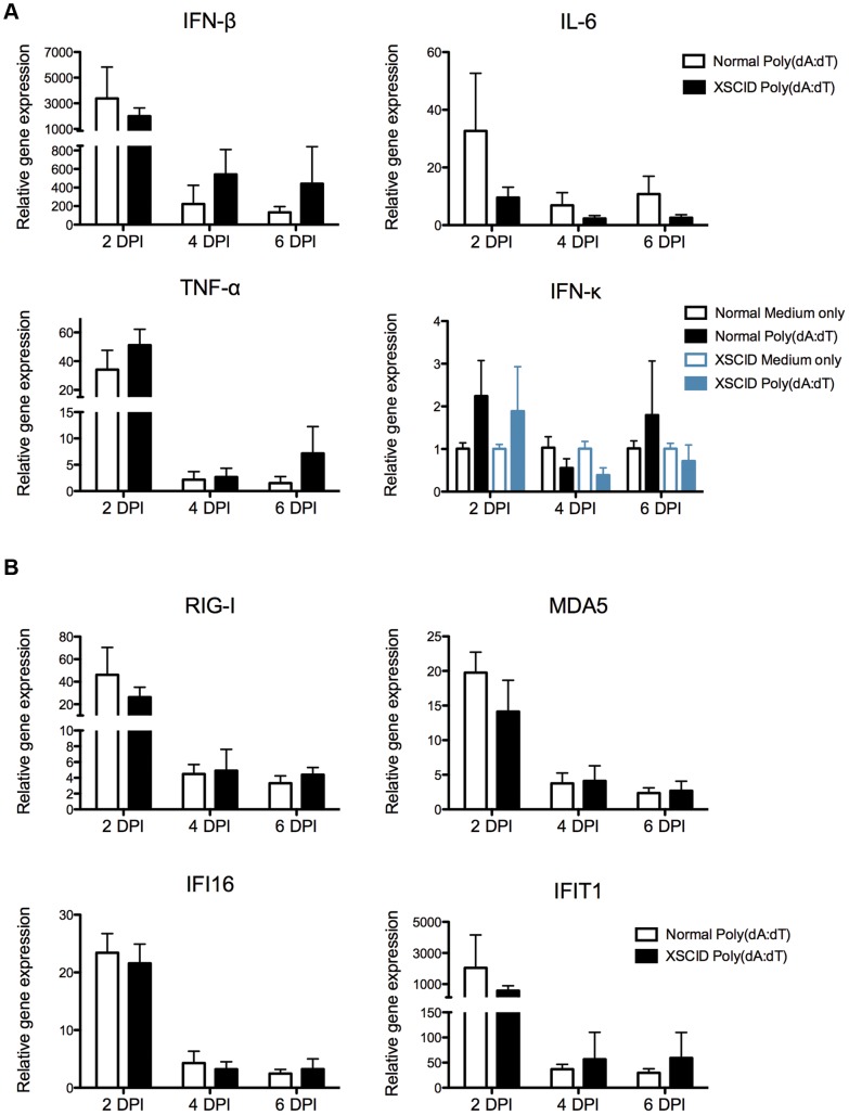
Figure 3. Kinetics of cytokine and interferon stimulated gene expression in XSCID canine keratinocytes stimulated with poly(dA:dT).
A and B Keratinocytes were seeded into multiple wells and cultured as a monolayer for 24(dA:dT) or medium alone. RNA was extracted after 2, 4, and 6 days post-stimulation. Cytokine (A) and interferon stimulated gene (B) expression was determined by quantitative RT-PCR. Resulting Cq values were normalized to a reference gene and calibrated to mRNA expression in unstimulated keratinocytes (ΔΔCq). Results are expressed as mean +/− SD of three replicate experiments performed in triplicate. Each experiment used keratinocytes derived from a different normal control dog (n = 3) and one of two different XSCID dogs (n = 2).

