Fig. 2.
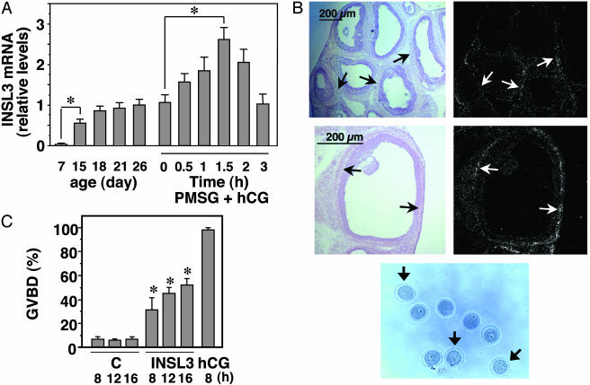
Regulation of thecal INSL3 expression and INSL3 induction of oocyte maturation in vivo.(A) Increases in ovarian INSL3 mRNA levels during development and after gonadotropin treatment. Quantitative RT-PCR was performed by using ovarian samples from rats at different ages (postnatal day). Ovaries were also collected before (day 26) and after treatment with 15 units of PMSG for 2 days (0 h of the PMSG + hCG group), followed by administration of 10 units of hCG for different intervals. The ratio of INSL3/β-actin transcript levels at 26 days of age was set as 1. *, P < 0.01. (B) In situ hybridization analysis of INSL3 expression in theca cells (arrows) of preovulatory follicles at 1.5 h after hCG treatment of PMSG-primed rats. (C) In vivo induction of oocyte maturation after INSL3 treatment. Immature rats at 2 days after PMSG priming were treated with INSL3 (2 μg per 0.1 ml of PBS) via intrabursal injections or hCG via s.c. injections. Oocytes were retrieved at different intervals after puncture of the ovary to release cumulus-oocyte complexes for assessing morphology. C, controls. *, P < 0.01 between control and INSL3-treated groups at the same time point. (D) Morphology of oocytes after INSL3 treatment (8 h) in vivo. Arrows indicate mature oocytes.

