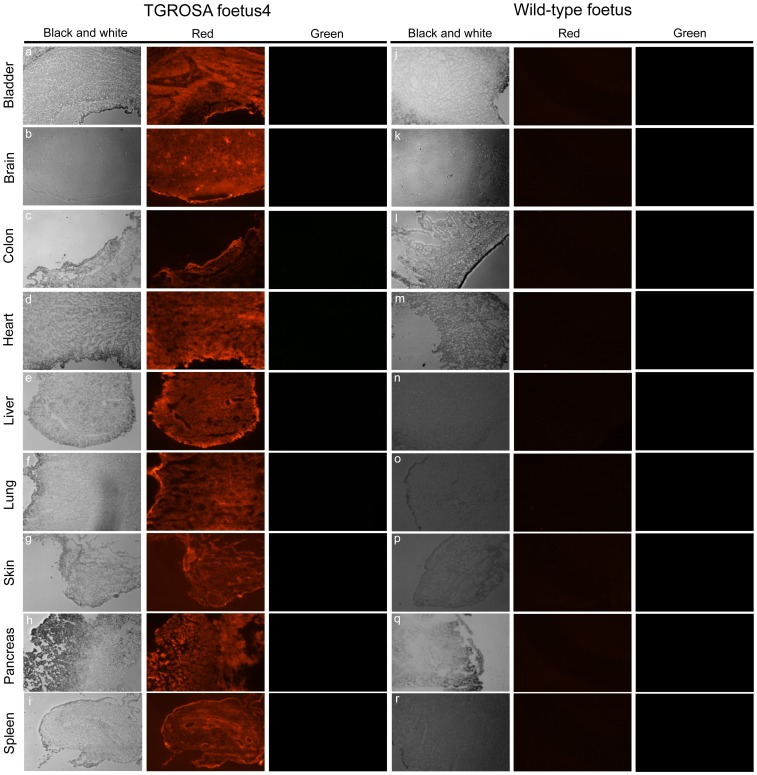
Figure 3. Fluorescence microscopy of tissue cryosections.
Panels a-i, organs from a nuclear transfer derived foetus as indicated. Panels j-r, organs from a wild-type foetus. Each section is visualised through black and white, red (excitation = 554 nm, emission = 581 nm) and green (excitation = 489 nm, emission = 509 nm) channels as indicated.

