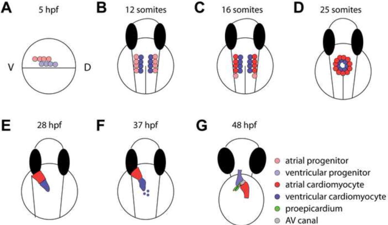
Figure 1.

Zebrafish heart developmental stages. (A) Fate mapping experiments identified cardiac progenitors in the lateral marginal zones on either side of the gastrula at ~5 hours post fertilization (hpf), with ventricular progenitors located more dorsally, and closer to the margin than atrial progenitors. (B, C) Cardiac progenitor cells continue to migrate towards the midline into the anterior lateral plate mesoderm (ALPM). From there, (B) the ventricular cardiomyocytes differentiate at the 12 somite stage (~16 hpf) as detected by vmhc expression, whereas (C) the atrial cardiomyocytes differentiate later at the 15 somite stage as detected by amhc/myh6 expression. During 19–26 hpf, additional differentiating atrial cardiomyocytes are added to the venous pole. (D) The progenitor cells further migrate to the midline to form a cardiac disc around the 25 somite stage (~22 hpf), where ventricular and atrial cardiomyocytes are located in the medial and peripheral regions of the disc, respectively. (E) As the cardiac tube extends and turns, the arterial pole is anterior and right-sided, whereas the venous pole is more posterior and left-sided. (F) Late differentiating cardiomyocytes add to the arterial pole from 34–48 hpf, and the heart tube begins to constrict at the future AV canal and loop at ~37 hpf. (G) By 48 hpf, the cardiac chambers have undergone ballooning/expansion, while the AV canal remains constricted. The first proepicardial cells can be observed at this stage near the ventral surface of the looped heart, next to the AV canal. These cells will proliferate and migrate to become the epicardium, which eventually covers the myocardium.
