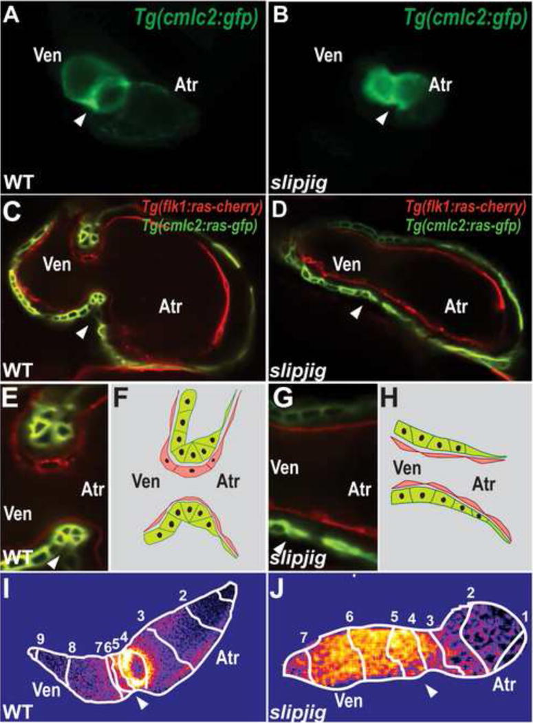
Figure 4.

slipjig/foxn4 mutant fails to form the AV canal and develop an AV conduction delay. (A, B) Epifluorescence micrographs show that Tg(cmlc2:GFP) (A) wildtype (WT) hearts loop by 48 hpf but (B) slipjig/foxn4 mutant hearts do not. (C, E) WT and (D, G) slipjig Tg(flk1:ras-cherry); Tg(cmlc2:ras-GFP) hearts, which have endocardial and myocardial cells outlined in red and green, respectively, were imaged by confocal microscopy at 40 hpf. AV myocardial and endocardial cell morphologies become triangular and cuboidal, respectively in (C, E) WT but not (D, G) slipjig/foxn4 hearts. These cell morphology changes are further illustrated in (F, H). (I, J) Optical mapping by calcium imaging using Tg(cmlc2:gCaMP) shows that AV conduction delay as observed in (I) WT is absent in (J) the slipjig mutant. Isochronal lines (numbered white lines) indicate temporal calcium activation every 60 ms from venous to arterial pole. White arrowhead: AV canal; Atr: atrium; Ven: ventricle. Numbers: calcium activation sequence. [39]
