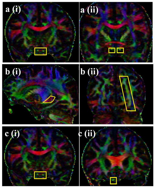
Figure 1.
The selected ROI seeds (ROI 1 and ROI 2) for optic tract and optic radiation are illustrated on the coronal DTI color-coded map. To trace the OT, the first ROI was placed over the optic chiasm in the coronal plane (1ai). The second ROI was pursued by selecting the anterior-posterior oriented green association fibers shown in the coronal plane passing through the level of the anterior commissure (Fig. 1aii). The first ROI to trace the OR was seeded on the red fibers of the thalamus (1bi) (right to left oriented fibers that loop over the temporal horn of lateral ventricle before turning medially toward the calcarine sulcus in the occipital lobe). The second ROI was pursued on the green fibers (anterior-posteriorly oriented fibers) in the occipital cortex (Fig. 1bii). The ROIs for delineation of the optic nerve are seen in Fig. 1ci and 1 cii. The first ROI was placed over the optic chiasm in the coronal plane (1ci). The second ROI was placed on the optic nerve at the level of the rostrum of the corpus collosum on the coronal plane.

