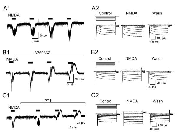
Fig. 1.
AMPK activators augment the ability of NMDA (10 μM) to evoke outward currents in STN neurons. (A1) Current trace shows that repeated applications of NMDA (10 μM) consistently evoke inward currents (at – 70 mV) in an STN neuron. Truncated deflections in these and subsequent current records are artifacts caused by voltage steps that were used to measure series resistance or membrane conductance for the construction of I-V plots. (A2) Current traces recorded during a series of hyperpolarizing voltage steps (from −70 to −140 mV) in the absence and presence of NMDA. Wash indicates recording after NMDA was washed from the slice. Dashed line indicates zero current. (B1) Current trace shows that NMDA evokes outward currents with increasing amplitudes when the slice is superfused with the AMPK activator A769662 (10 μM). (B2) Current traces recorded during a series of hyperpolarizing voltage steps (from −70 to −140 mV) show that NMDA increases membrane conductance in an STN neuron when recorded in the presence of A769662 (10 μM). (C1) Current trace shows that NMDA evokes outward currents with increasing amplitudes when the slice is superfused with the AMPK activator PT1 (10 μM). (C2) Current traces recorded during a series of hyperpolarizing voltage steps (from −70 to −140 mV) show that NMDA increases membrane conductance in an STN neuron when recorded in the presence of PT1 (10 μM).

