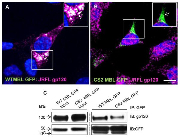
Figure 2. MBL interacts and co-localizes with JRFL gp120 in perinuclear vesicles.
Confocal laser scan microscopy analysis of subcellular localization of WT MBL-EGFP (A) or Mut CS2 MBL-EGFP (B) with JRFL gp120. SK-N-SH cells for gp120 (magenta) followed by corresponding secondary antibodies and MBL WT and CS2 constructs were visualized by fluorescence of GFP (green). WT MBL-EGFP (closed arrows), (A) but not Mut CS2 MBL EGFP (open arrows) (B) co-localized with JRFL gp120 at perinuclear vesicles. White indicates co-localization. DAPI (blue) stained the nuclei. Scale bar, 10 μm. C, CRD-dependent subcellular interaction of MBL with gp120. SK-N-SH cells were co-transfected with WT MBL-EGFP or CS2 MBL-EGFP and JRFL gp120 plasmids with Mirus Trans IT-LT1. 36h post-transfection cells were solubilized with 1% NP40 lysis buffer in the presence of Ca2+ and immunoprecipitated with anti-GFP polyclonal antibody. The immunoblots were probed with antibodies against gp120 and GFP respectively.

