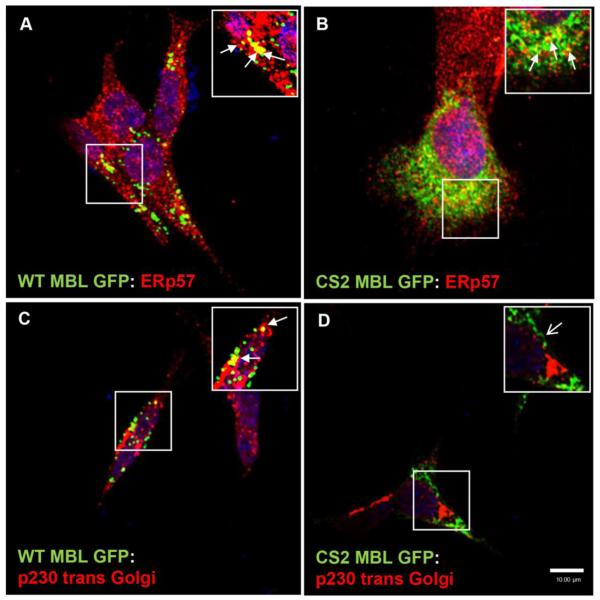
Figure 3. MBL accumulates and localizes in ER and Golgi vesicles.
A and C, Confocal laser scanning microscopy analysis of the subcellular co-localization of WT MBL EGFP with ER marker (ERp57) and Golgi marker (p230 trans Golgi), (closed arrows; yellow indicates co-localization) in SK-N-SH cells. B, Confocal laser scanning microscopic analysis of subcellular co-localization of CS2 MBL EGFP with ER marker (closed arrows; yellow indicates co-localization). D, Diffused distribution of CS2 MBL EGFP in the cytoplasm and absence of CS2 MBL-EGFP localization in Golgi (open arrow). WT and CS2 MBL-EGFP (green) expressing cells were stained with an ER marker, Erp57 (red) or Golgi marker p230 trans Golgi (red), followed by corresponding Alexa Fluor secondary antibodies. DAPI (blue) stained nuclei. Scale bar, 10 μm.

