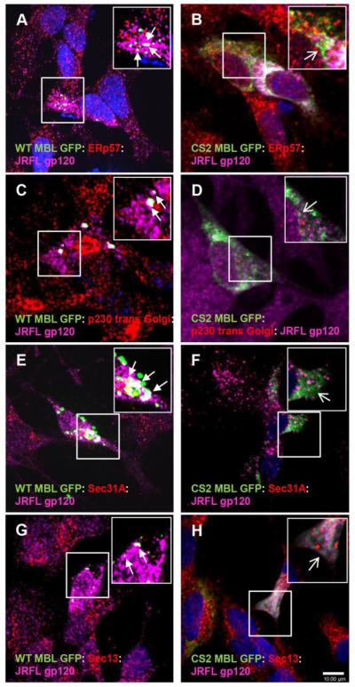
Figure 4. Functional co-localization of MBL:gp120 complexes with COPII transport vesicles markers.
A,C,E,G Functional co-localization of WT MBL EGFP (green) and JRFL gp120 (magenta) with ER marker (red), Golgi marker (red) and COPII transport vesicles markers (red): Sec 31A and Sec 13 (closed arrows, white indicates co-localization of three markers) in SK-N-SH cells. B, Localization of CS2 MBL (green) and JRFL gp120 (magenta) in ER (red) (open arrows). D, F, H Localization of CS2 MBL EGFP (green) and JRFL gp120 (magenta) with COP II transport vesicles: Sec 31A or Sec 13 (red) (open arrows). DAPI (blue) stained nuclei. Scale bar, 10 μm.

