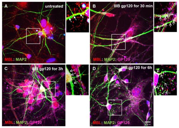
Figure 6. IIIB gp120 co-localizes with MBL and MAP2 and is transported along the microtubules in human primary neurons.
Human primary neurons were untreated (A) or treated with IIIB gp120 (5 nM) for 30min (B), 3h (C) and 6h (D). Neurons were fixed and stained for MBL (red), MAP2 (green), gp120 (magenta) and DAPI for nuclei (blue). Localization of MBL:MAP2 (closed arrows; yellow indicates co-localization) and MBL:MAP2:gp120 complexes (open arrows; white indicates co-localization) in neuronal soma and neurites. Dashed open arrows indicate fragmentation of the microtubules (D). Scale bar, 10 μm.

