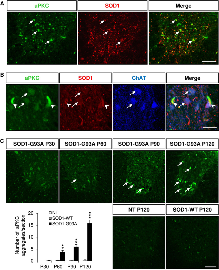
Figure 3. aPKC is colocalized with human SOD1 in extracellular aggregates.
Immunostaining of coronal sections of lumbar spinal cords of non transgenic (NT), SOD1 (WT) and SOD1 (G93A) mice of 120 days for aPKC (green), human SOD1 (red) and ChAT (blue). A, aPKC colocalizes with human SOD1 in the lumbar spinal cord of P120 SOD1 (G93A) mice (arrows). B, Higher magnifications showing colocalization of aPKC with human SOD1 protein in ChAT+ motor neurons (arrows) and in extracellular aggregates (arrowheads) in the ventral lumbar spinal cord of P120 SOD1-G93A mice. C, Quantification of the number of aPKC aggregates per section in NT, SOD1 (WT) and SOD1 (G93A) mice at different stages. Data represent mean±SEM, n=3–5 animals/group, ** p<0.01, *** p<0.001, ANOVA with Bonferroni Post-test. Scale bars: 50 µm (A, C), 25 µm (B).

