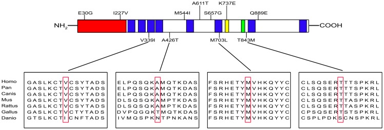
Figure 2. Locations of mutations identified and sequence alignments in ZFPM2/FOG2.
Top, the N-terminal transcriptional repression domain is indicated in red; the eight zinc-finger motifs are represented by blue; the nuclear localization signal is indicated in yellow; the putative CtBP-binding site is represented by green. Variants identified in this study are shown underneath the protein structure; variants reported in previous studies are shown above. Bottom, multiple alignment of partial amino acid sequences of human ZFPM2/FOG2 and its homologs from other species. Variant residues are boxed. Accession numbers of the sequences used are as follows: Homo, NP_036214.2; Pan, XP_001158075.1; Canis, XP_539118.2; Mus, NP_035896.1; Rattus, XP_235253.4; Gallus, XP_418380.2; Danio, NP_001034724.1.

