Fig. 1.
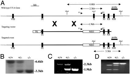
Targeted disruption of the mouse UT-A gene. (A) Structure of the wild-type UT-A gene, the targeting vector, and the mutant allele. Exons are represented as filled boxes, and unique restriction enzyme sites are shown. The expression cassettes for the neomycin resistance gene (NEO) and thymidine kinase (TK) gene are shown as open boxes. Position of external probe and the length of restriction fragments and primers used for genotyping are denoted by arrows and arrowheads, respectively. (B) Representative Southern blot analysis of genomic DNA. XbaI-digested DNAs from wild-type, heterozygous, and UT-A1/3-/- mice were hybridized with probe as shown in A. Sizes (in kb) of bands are shown. (C) Representative results of PCR genotyping with primers (F1 and R1) as shown in A. Sizes (in kb) of bands are shown. (D) Restriction digestion of aforementioned PCR products with AccI, unique to exon 10, results in the complete digestion of the wild-type PCR product, but not the knockout allele.

