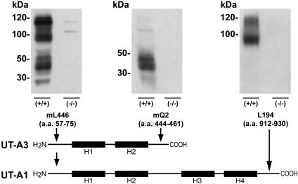
Fig. 2.
Representative immunoblots of whole IM homogenates probed with several isoform-selective polyclonal antibodies. The schematic diagram shows the antibodies used and the protein isoforms they detected. H1-H4 shows putative membrane-spanning regions. When using antibody mL446, strong protein bands of ≈100 and 120 kDa (glycosylated forms of UT-A1) and a protein “smear” between 40 and 56 kDa (glycosylated UT-A3) are absent from UT-A1/3-/- mice. When using antibody mQ2, which selectively recognizes UT-A3, a protein smear between 40 and 56 kDa is absent in UT-A1/3-/- mice. When using antibody L194, strong protein bands of ≈100 and 120 kDa (UT-A1) are absent in UT-A1/3-/- mice.

