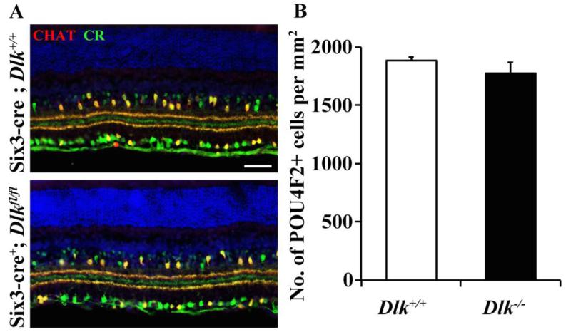
Figure 1. Dlk deficiency does not appear to significantly affect RGC development.
(A) Representative images from wildtype and Dlk deficient (Six3-cre+; Dlkfl/fl) retinas. Deficiency in Dlk throughout retinal development (Six3-cre) did not alter the gross lamination in the inner plexiform layer, as judged by normal organization of calretinin (CR, green) and choline acetyl transferase (CHAT, red) processes (3 retinas of each genotype were assessed). (B) Cell counts of POU4F2 labeled RGCs in the adult retina show that embryonic deletion of Dlk in the retina does not alter developmental cell death of RGCs (P = 0.316; N=6 for each genotype). Scale bar: A, 50 μm.

