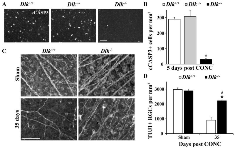
Figure 2. Dlk deficiency delays RGC death after axonal injury.
(A) Representative images from Dlk+/+, Dlk+/- and Dlk−/− flat mounted retinas stained with anti-cleaved caspase 3 (cCASP3) 5 days after CONC, a time point where RGC death peaks (Harder et al., 2012). (B) Cell counts of cCASP3 positive cells at 5 days after CONC (N ≥ 6 for each genotype). Complete deficiency in Dlk significantly lessened the number of dying RGCs after CONC compared to wildtype retinas (P < 0.001). The number of dying cells was similar in Dlk+/- mice compared to wildtype retinas (P = 0.61). To determine if deficiency in Dlk affected the long term survival of RGCs after axonal injury, the number of surviving RGCs (TUJ1+ cells) were counted at 35 days after CONC, a time point when the majority of RGCs have died in wildtype retinas (C,D). (C) Representative images from Dlk+/+ and Dlk−/− flat mounted retinas stained for the RGC marker TUJ1 35 days after CONC. (D) TUJ1 cell counts show that significantly more RGCs survive 35 days after CONC in Dlk deficient retinas compared to wildtype retinas (* P < 0.001). However, at 35 days after CONC, there is significant decrease in RGC numbers in Dlk deficient retinas compared to sham-injured retinas (# P < 0.05). N ≥ 4 for each genotype and condition (sham, CONC); scale bar: A, C, 50 μm.

