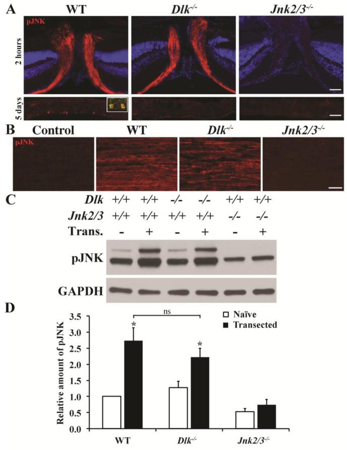
Figure 3. Dlk deficiency does not prevent JNK activation in RGC axons after CONC.
(A) Immunohistochemistry of retinal sections containing the optic nerve head (ONH) 2 hours after CONC shows robust pJNK staining in RGC axons of wildtype mice. Dlk deficiency did not prevent JNK activation in RGC axons in the ONH. As previously shown, combined deficiency of Jnk2 and Jnk3 prevents pJNK detection in RGC axons (Fernandes et al., 2012). Five days after CONC robust pJNK staining is detected in RGC somas in wildtype retinas (inset shows pJNK expressed in a TUJ1+ RGC). Interestingly, despite the presence of pJNK in RGC axons in Dlk deficient mice, pJNK does not appear to be present in somas 5 days after CONC. pJNK was also not present in RGC somas in mice deficient in both Jnk2 and Jnk3. (B) To determine if Dlk is required to phosphorylate JNK in axons, optic nerves were transected both at the retinal and chiasm ends and the optic nerve segments were cultured. Optic nerve transection induces robust activation of JNK in wildtype nerves. In Dlk deficient nerves, pJNK was still detected indicating that Dlk is not required for activation of the axonal pool of JNK in RGCs following axonal injury. (C) Representative western blots showing levels of pJNK increase in optic nerve segments that are transected and cultured for 4 hours. (D) There was a significant increase in pJNK levels in both wildtype and Dlk deficient optic nerves but not Jnk2/3 deficient optic nerves after transection. Additionally, pJNK levels after transection were not significantly different between wildtype and Dlk deficient optic nerves. At least 3 retinas or optic nerves were assessed for each genotype and condition. *, P < 0.05; ns, not significant; Scale bar: A, 50 μm; B, 20μm.

