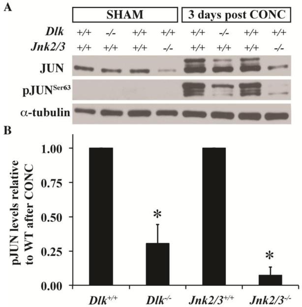
Figure 4. Activation of JUN is attenuated in the Dlk deficient mice.
(A) Representative western blot showing the expression of JUN and pJUNSer63 in uninjured (Sham) retinas and in retinas 3 days post controlled optic nerve crush (CONC). Note that the Ser63 residue of JUN is phosphorylated by JNK. Levels of JUN in uninjured retinas appear to be similar in wildtype and Dlk deficient retinas. In Jnk2/3 deficient mice, JUN levels are less than in wildtype retinas, suggesting that JNK2 and/or 3 participate in regulating the physiological levels of JUN in uninjured Sham retinas. There is no detectable level of pJUNSer63 in uninjured retinas of any genotype (data not shown). Three days after CONC, JNK phosphorylation of JUN occurs in wildtype retinas. Levels of pJUNSer63 are reduced in Dlk deficient and Jnk2/3 deficient retinas. Interestingly, most of the JUN that is phosphorylated in Dlk deficient retinas appears to be phosphorylated at multiple sites as suggested by predominance of the higher molecular weight band in contrast to predominant lower molecular weight band observed in Jnk2/3 deficient retinas. (B) Quantitation of the western data shows that deficiency in Dlk or Jnk2/3 significantly reduces JUN phosphorylation 3 days after CONC. Note, the amount of pJUN 3 days after CONC is normalized to the loading control and is expressed relative to the injured wildtype. N = 3 for all conditions and genotypes. *, P < 0.05.

