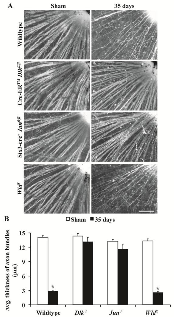
Figure 5. Axons do not degenerate proximal to injury site in Dlk deficient mice.
(A) Representative images adjacent to the optic nerve head of retinal flatmounts (RGC layer up) stained with the RGC marker, TUJ1. Dlk deficiency and Jun deficiency have similar numbers of surviving RGCs 35 days after injury (see above, Dlk−/−, 76.9%; Jun−/−, 69.3%; Dlk and Jun floxed alleles were recombined with Cre-ER™ and Six3-cre respectively). In Dlk deficient retinas, numerous RGC axons were present on the surface of the retina, suggesting that Dlk has a role in axonal degeneration. However, the axon bundle thickness appeared similar in Jun deficient retinas, suggesting that proximal RGC axonal survival is linked to somal survival. In contrast, there appeared to be extensive loss of proximal axon segments in WldS retinas, which do not have significant somal survival compared to wildtype retinas at this time point (Wang et al., 2013a). (B) Quantitative analysis of proximal axon bundle thickness shows that the proximal axon degenerates in both WT and WldS retinas 35 days after CONC. However, there is no significant reduction in the thickness of the axon bundle in either Dlk−/− or Jun−/− retinas at 35 days post CONC. At least 3 retinas were examined for each genotype and condition. *, P < 0.05; Scale bar, 100 μm.

