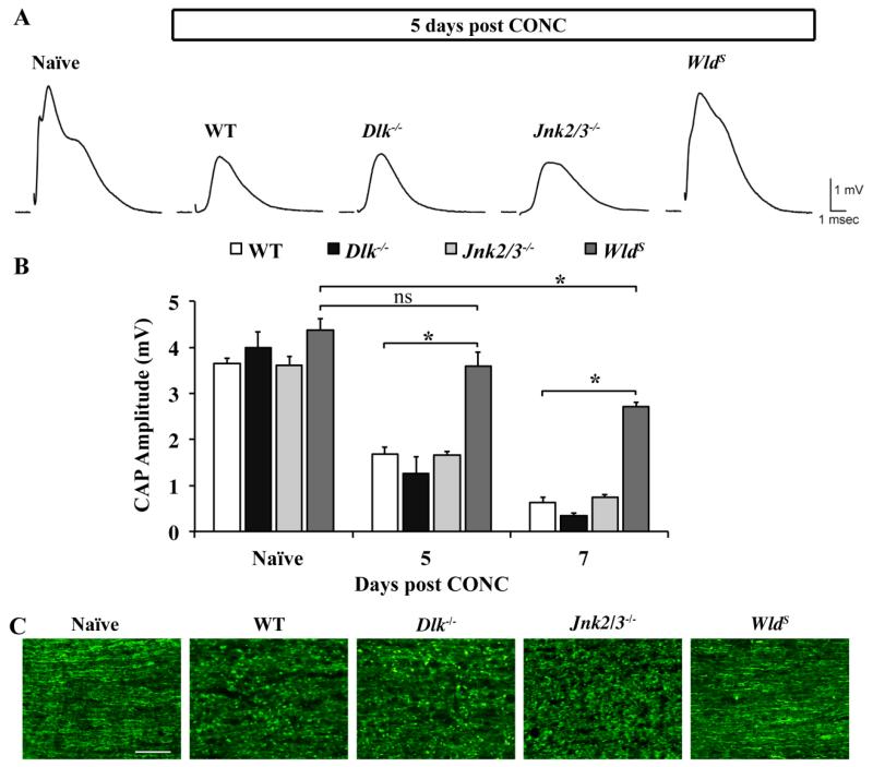
Figure 6. Dlk deficiency does not appear to affect distal axonal degeneration.
To determine if Dlk was important in axonal degeneration distal to the site of injury, compound action potentials (CAP) were recorded from wildtype, Dlk−/−,, Jnk2/3−/−, and WldS optic nerves at 5 or 7 days after CONC. Dlk or Jnk2/3 deficiency did not rescue the reduction in amplitude of the CAP observed after CONC and both were significantly reduced compared to naïve control mice (P < 0.001 for each time point and genotype). As expected, optic nerves from WldS mice were protected from axon degeneration following CONC. The CAP amplitude of WldS optic nerves 5 days after CONC was not significantly different from the naïve control. Seven days after CONC there was a significant reduction in the CAP amplitude in WldS nerves compared to uninjured naïve control (P = 0.006). However, at both 5 and 7 days after CONC, the CAP of WldS nerves was significantly higher than wild-type nerves (P<0.001). (C) Representative images of longitudinal optic nerve sections show that there was extensive axonal fragmentation and beading in wildtype, Dlk−/− and Jnk2/3−/− optic nerves after CONC. However, optic nerves from WldS mice 5 days after CONC appeared intact, supporting the functional rescue observed in WldS mice at this time point. N ≥ 3 for each genotype and condition (sham, CONC). *, P < 0.05; ns, not significant; Scale bar: 50 μm.

