
Figure 3.
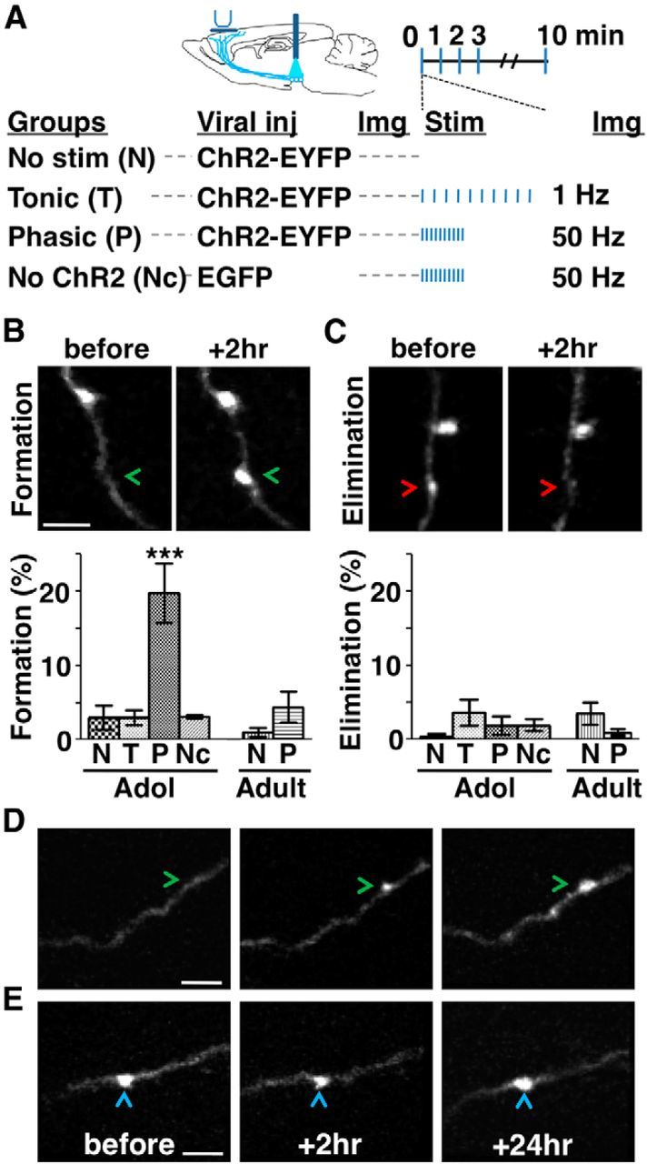
Phasic activation promotes mesofrontal bouton formation in adolescence. A, Experimental conditions. B, C, Top, two-photon images showing bouton formation (B, arrowheads) and elimination (C, arrowheads) on mesofrontal axons over a 2 h interval. Bottom, Percentage of boutons formed (B) and eliminated (C) in adolescent (∼4–5 weeks) and adult (∼2–4 months) male mice. For B, one-way ANOVA F(5,30) = 12.09, p < 0.0001; Bonferroni's posttest, ***p < 0.001, adolescent phasic group versus all other groups. For C, one-way ANOVA F(5,30) = 1.33, p = 0.279. n = 6 mice each group. Data are means ± SE. D, E, Two-photon images showing newly formed (D, arrowheads) and preexisting (E, arrowheads) boutons identified 2 h after phasic stimulation persisted after 24 h in adolescent mice (repeated in 9 mice). The majority (79%, 19/24) of new axonal boutons formed 2 h after phasic stimulation were still present 24 h later, comparable to the stability of boutons that existed before stimulation (88%, 109/124; p = 0.75, χ2 test). Scale bars, 5 μm.