
Figure 4.
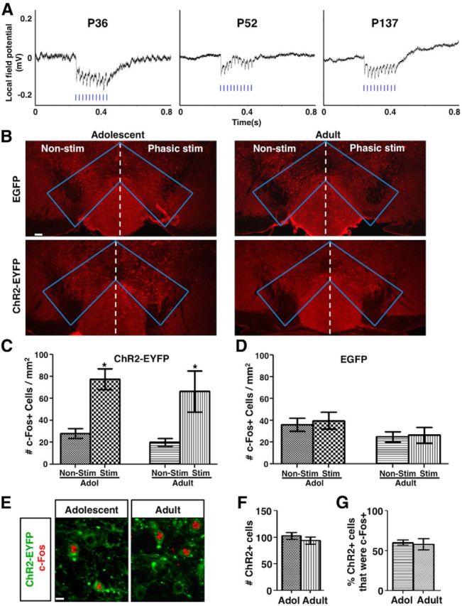
Phasic activation induces robust LFP response and comparable c-Fos expression in adolescent and adult mice. A, VTA LFP traces in response to phasic light stimulation of ChR2-expressing dopamine neurons in mice with ages ranging from adolescence to adulthood. 50 Hz LFP responses time locked to 50 Hz phasic light pulses (blue bars) were observed in all ages. Average of 10 sweeps of LFP recordings are shown for each condition. B, Confocal images of c-Fos immunofluorescence in the VTA of adolescent (∼4–5 weeks) and adult (∼2–4 months) mice that were transduced with either AAV:ChR2-EYFP or AAV:EGFP and unilaterally stimulated with phasic light pulses. C, In AAV:ChR2-EYFP-transduced mice, the density of c-Fos+ cells at the stimulated side was increased to a similar extent between adolescent (∼4–5 weeks) and adult (∼2–4 months) mice. RM-ANOVA: stimulation, F(1,6) = 27.80, p = 0.002; age, F(1,6) = 0.585, p = 0.474; simulation-by-age interaction, F(1,6) = 0.034, p = 0.859; nonstimulated versus stimulated, adolescent *p = 0.017, adult *p = 0.023, Bonferroni's posttests, n = 4 mice for each group. D, In AAV:GFP-transduced mice, no significant difference was observed. RM-ANOVA: stimulation, F(1,6) = 0.990, p = 0.358; age, F(1,6) = 1.91, p = 0.216; interaction, F(1,6) = 0.196, p = 0.674. n = 4 mice for each group. E, Confocal images of ChR2-EYFP (green) and c-Fos (red) labeled cells in the VTA of adolescent and adult mice after phasic stimulation. F, G, The number of ChR2+ cells (F) in 636 × 636 × 12 μm3 image stacks from two sections/mouse and the percentage of ChR2+ cells that were c-Fos+ (G) are not different between adolescent and adult mice. p = 0.390 (F) and 0.789 (G), t tests, n = 4 mice each. Scale bars: B, 100 μm; E, 10 μm. Data are means ± SE.