
Figure 1.
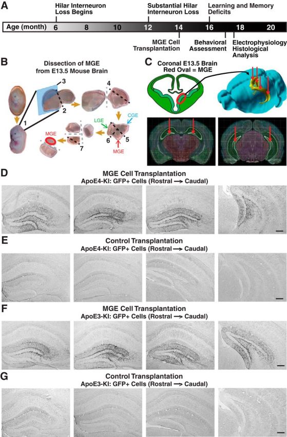
Experimental timeline and protocol, hilar targeting, and migration of transplanted MGE cells in apoE4-KI and apoE3-KI mice. A, Experimental timeline for hilar MGE cell transplantation and evaluation. B, Dissection protocol of MGE from GFP+ E13.5 mouse embryos. (1) Removal of embryonic brain, (2) isolation of the telencephalon by removing hindbrain, (3) sagittal cut to separate two hemispheres, (4) isolation of ventral telencephalon by removing dorsal cortex, (5) removal of caudal ganglionic eminence (CGE) and (6) lateral ganglionic eminence (LGE), and (7) removal of the mantle zone and preoptic area for dorsal MGE collection. C, The dissected MGE cells from GFP+ E13.5 mouse embryos were bilaterally transplanted into the rostral and caudal hilus of 14-month-old apoE4-KI mice. D–G, Immunostaining of GFP+ cells in hippocampal sections 1.2 mm apart from living MGE cell-transplanted apoE4-KI mice (D), control-transplanted apoE4-KI mice (E), living MGE cell-transplanted apoE3-KI mice (F), and control-transplanted apoE3-KI mice (G) at 80–90 DAT. Scale bars: D–G, 250 μm.