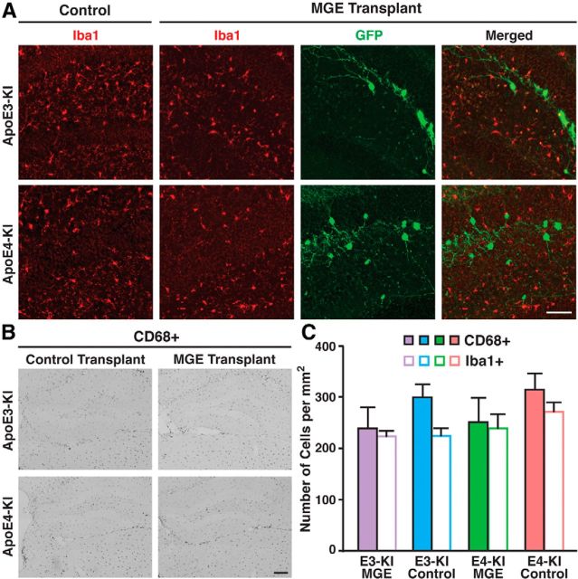
Figure 5.
Examination of microglia as a marker for inflammation. A, Immunofluorescent staining of microglial marker Iba1 showed no microglia clustering around the grafted cells. B, Immunostaining of activated microglia marker CD68 showed no significant differences between MGE cell-transplanted and control mice. C, Quantification of CD68+ and Iba1+ cells per square millimeter (n = 5–8 sections per brain, 3–5 mice per group). Values are shown as the mean ± SEM. Scale bars: A, 50 μm; B, 250 μm.

