
Figure 6.
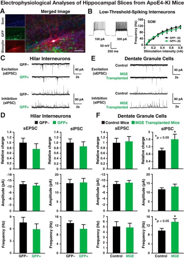
Electrophysiological analyses of transplanted and endogenous cells in acute hippocampal slices from 17-month-old apoE4-KI mice at 80–90 DAT. A, Post hoc staining of a low-threshold-spiking GFP+ interneuron filled with biocytin during recording was positive for SOM. Scale bar, 50 μm. B, Representative voltage traces from a GFP+ hilar cell are shown. The intrinsic excitability study (frequency of firing vs stimulation intensity) of GFP+ hilar cells revealed firing properties indistinguishable from endogenous (GFP−) hilar cells. C, D, Transplanted GFP+ cells displayed spontaneous EPSCs (sEPSCs) and spontaneous IPSCs (sIPSCs; C) at frequencies and amplitudes (D) comparable to endogenous (GFP−) hilar interneurons. E, F, Current traces (E) from dentate granule neurons in control and MGE cell-transplanted apoE4-KI mice show no quantifiable changes (F) in sEPSCs, but a significant increase in the frequency of sIPSCs, leading to an overall increase in inhibitory charge transfer. Values are shown as the mean ± SEM. *p < 0.05, t test. n = 8–15 cells per group.