
Figure 7.
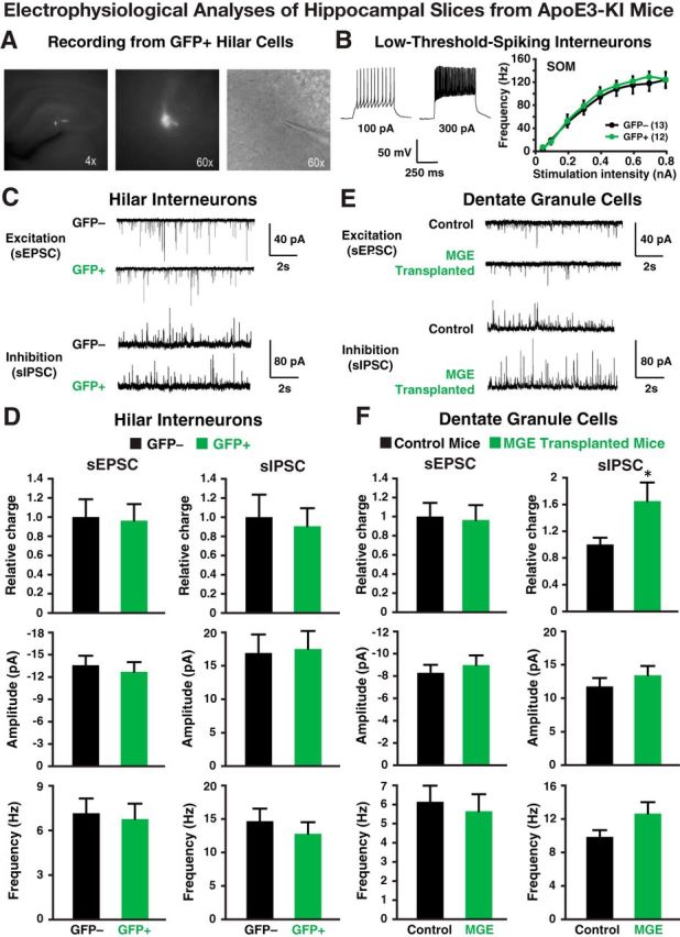
Electrophysiological analyses of transplanted and endogenous cells in acute hippocampal slices from 17-month-old apoE3-KI mice at 80–90 DAT. A, Fluorescent and bright-field images (4× and 60×) of a GFP+ hilar cell during electrophysiological recording. B, Representative voltage traces from a GFP+ hilar cell are shown. The intrinsic excitability study (frequency of firing vs stimulation intensity) of GFP+ hilar cells revealed firing properties indistinguishable from endogenous (GFP−) hilar cells. C, D, Transplanted GFP+ cells received spontaneous EPSCs (sEPSCs) and spontaneous IPSCs (sIPSCs; C) from the host cells at frequencies and amplitudes (D) comparable to endogenous hilar interneurons (GFP−). E, F, Current traces (E) from dentate granule neurons in control and MGE cell-transplanted apoE3-KI mice reveal no quantifiable changes (F) in excitatory input (i.e., sEPSCs), but a significant increase in inhibitory charge transfer. Values are shown as the mean ± SEM. *p < 0.05, t test. n = 8–15 cells per group.