Figure 2.
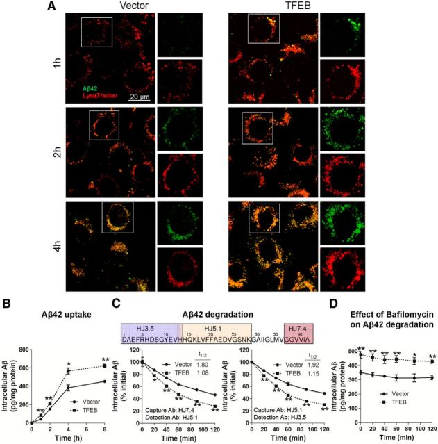
TFEB enhances Aβ uptake and degradation in C17.2 cells. A, Representative confocal images of C17.2 cells transfected with TFEB or empty vector, and incubated with 500 nm FAM-Aβ42 at varying times, as indicated and imaged with LysoTracker Red colabeling. Representative of n = 3 independent experiments. B, C17.2 cells were transfected with TFEB or empty vector for 48 h, and subsequently incubated with Aβ42 (500 nm) for an additional 1–8 h, and intracellular Aβ42 was analyzed by ELISA at the time points indicated. N = 3/group per time point. C, C17.2 cells were transfected with TFEB or empty vector for 48 h and Aβ42 (500 nm) was applied for 4 h, followed by its removal. Cells were then thoroughly washed. At varying times after washing (as indicated), the cells were trypsinized, lysed, and intracellular Aβ42 was quantified by ELISA using two separate strategies. Specific antibodies used (refer to schematic, top) are noted (bottom). N = 3/group per time point. D, Cells treated as in C with the addition of Bafilomycin A1 (100 nm) for 30 min before washing out the Aβ and cultured in its presence until the cells were collected for assay. N = 4/group; *p < 0.05, **p < 0.01.

