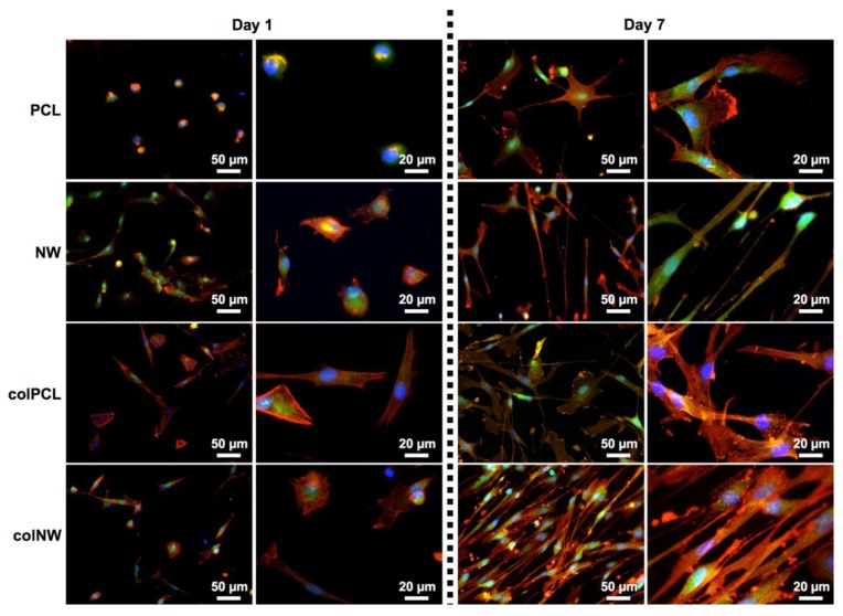
Figure 4.
Representative fluorescence microscopy images of SMCs stained with 5-chloromethylfluorescein diacetate (CMFDA) (green), rhodamine phalloidin (red) and DAPI (blue) on the PCL, NW, colPCL and colNW surfaces. Experiments were replicated on at least three different samples with at least three different cell populations (nmin = 9).

