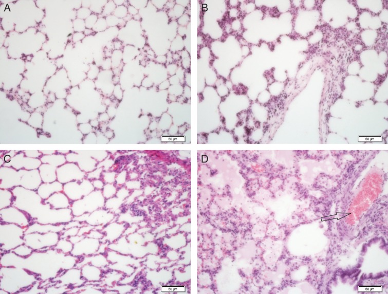
Fig. 1.
Pulmonary histology after irradiation. Lung sections from a sham-irradiated rat (A) and rats 7 d (B), 14 d (C), and 28 d (D) after irradiation of 17 Gy were stained with H&E. On Days 7 and 14 after irradiation, intra-alveolar edema was obvious due to increased vascular permeability and exudation of proteins into the alveolar space. The alveolar space was integrated, pulmonary capillaries were expanded, and hyperemia was present with some inflammatory cell infiltration (B, C). Enlarged inflammatory cell infiltration was observed on Day 28 after irradiation, consistent with pulmonary edema, vessel thrombosis (arrows), and intra-alveolar hemorrhage. Diffuse inflammatory cell infiltration was detected in broad areas of the lung parenchyma (D). Scale bar: 50 µm.

