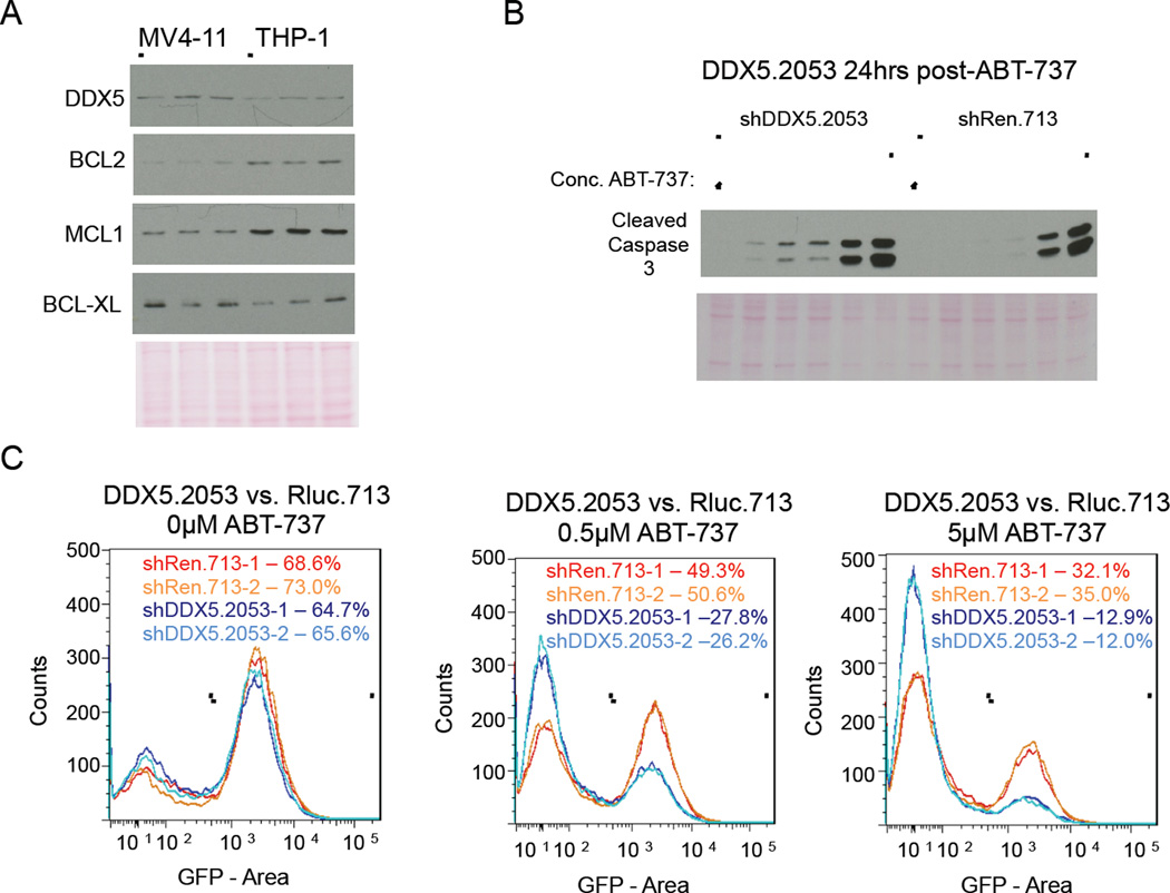
Figure 5. Combined inhibition of DDX5 and BCL2 family proteins cooperate to induce apoptosis in THP1 AML cells.
(A) Immuno-blot analysis of BCL2 family proteins in WCEs from the indicated AML cell lines. Note WCEs prepared from 50,000 cells per cell line were loaded onto the immuno-blots and results for triplicate independently prepared WCEs from each cell line are presented. (B) Immuno-blot analysis of Caspase 3 activation for THP1 cells +/− DDX5 knockdown treated for 24hrs with increasing concentration of ABT-737. Concentrations of ABT-737 tested were 0µM, 0.1µM, 0.5µM, 1µM, 5µM, and 10µM. (C) Flow cytometry analysis of live GFP positive cells in THP1 cultures +/− DDX5 knockdown untreated or treated for 24hrs with either low dose (0.5µM) or high dose (5µM) ABT-737. Results from duplicate cultures per RNAi condition are presented. GFP positive cells were gated and the percent of GFP positive cells in each culture are shown.

